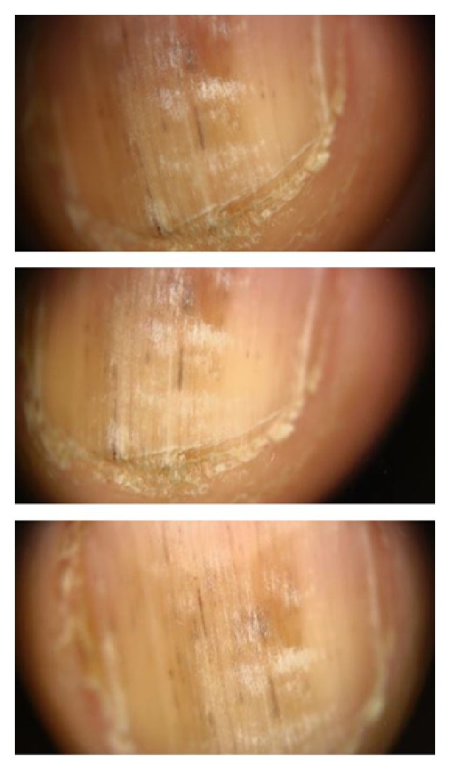
Figure 2.

Dermoscopic evaluation of right second nail yields longitudinal gray regular lines on a grayish background, splinter hemorrhages as blackish linear streaks, and true leukonychia as it did not disappear with pressure and hyperkeratosis in hyponychium.
