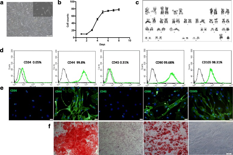
Fig. 1.

Identification of hMSCs. a Morphological appearance of third-passage hMSCs. Scale bar = 200 μm (inset 20 μm). b Logarithmic proliferation of cells. c Chromosome karyotype analysis of cells. d hMSC surface markers evaluated through flow cytometric analysis. e Immunofluorescence performed using monoclonal antibodies. f Differentiation of hMSCs into adipocytes and osteoblasts. Scale bar = 100 μm. n = 3
