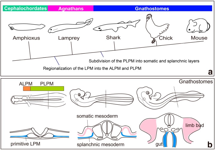
Figure 1.
Model for the evolution of the lateral plate mesoderm (LPM); (a) a phylogenetic tree indicating the probable timing of the regionalization and subdivision of the LPM. See text for details. Modified after [4]. (b) development of the LPM in chick embryos. Bottom panels show schematic cross-sections at the wing level. The LPM is regionalized into the anterior lateral plate mesoderm (ALPM) and the posterior lateral plate mesoderm (PLPM). Then, the developing LPM splits into somatic and splanchnic mesoderm. Subsequently, somatic mesodermal cells proliferate and form limb buds, whereas splanchnic mesodermal cells contribute to gut formation. Modified after [9].

