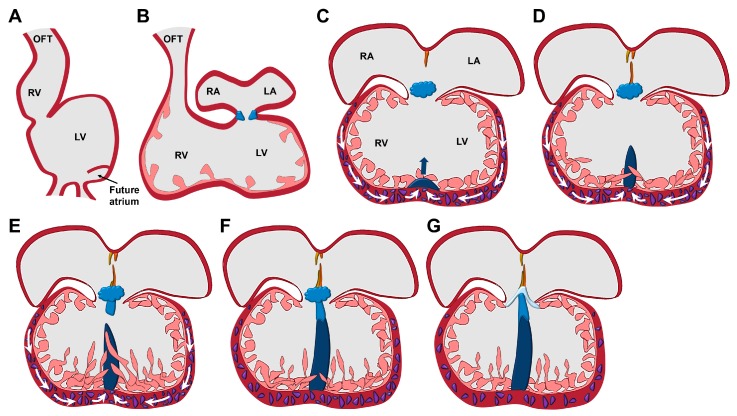
Figure 1.
Development of the ventricular septum. (A) The linear heart tube balloons to give rise to precursor structures of the heart chambers. (B) The heart takes its four-chambered shape by a process termed heart looping. (C) Proliferating cells (in purple) of the ventricular walls lead to the outgrowth of the muscular ventricular septum (in dark blue). (D) In addition, trabeculae that are derived from the ventricular walls start to participate in the formation of the ventricular septum. (E) After a molecular interaction between the muscular ventricular septum and the endocardial cushion cells (in bright blue), the membranous ventricular septum develops from the endocardial cushion cells and grows towards the muscular ventricular septum. (F) Finally, the muscular and membranous ventricular septa fuse. (G) The atrioventricular endocardial cushion cells give rise to the atrioventricular valves. LA, left atrium; LV, left ventricle; OFT, outflow tract; RA, right atrium; RV, right ventricle.

