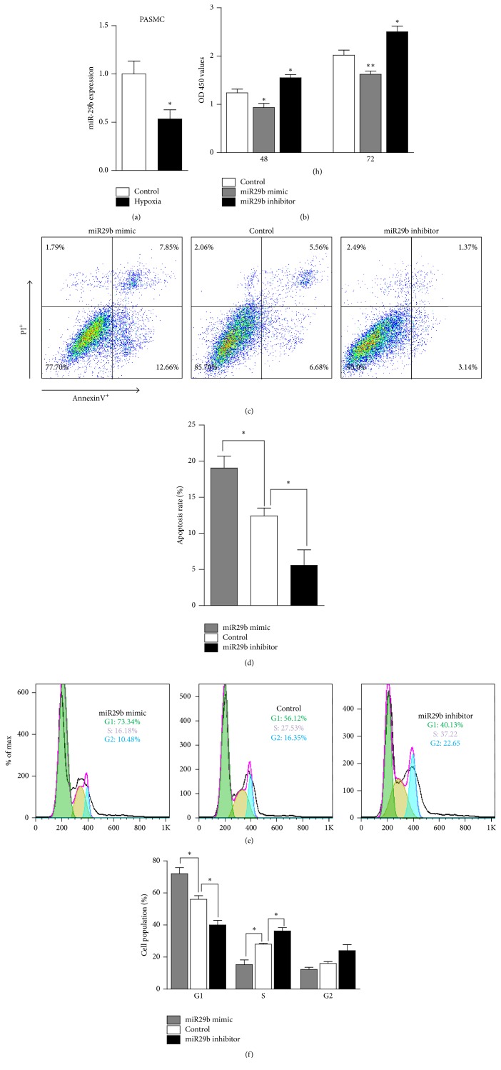
Figure 2.
MiR-29b promotes apoptosis of PASMCs. (a) The expression of miR-29b in PASMCs cultured under hypoxia or normal air (control) for 48 h (n = 6 mice/group). (b) The proliferation of PASMCs following transfection with the miR-29b mimic, inhibitor, or negative control for 48 h or 72 h analyzed using a CCK-8 assay kit. The OD450 value revealed the number of PASMCs (n = 6 mice/group). (c and d) The representative graph and statistical chart of the apoptosis rate of PASMCs treated as indicated for 48 h determined by flow cytometry following AnnexinV and PI staining. (e and f) The representative graph and statistical chart of the number of PASMCs treated with the miR-29b mimic, inhibitor, or negative control for 48 h in the G0/G1, S, and G2/M phases determined by flow cytometry after staining with PI. ∗p < 0.05 and ∗∗p < 0.01.

