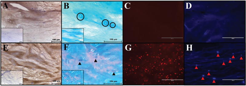
Figure 3.

Histological analysis of ABNP scaffolds (A–D) compared to fresh bovine NP tissue (E-H). (A&E) Immunohistochemistry for Col-II (brown = positive, light blue = nuclei) (Insert = negative control) (200×). (B&F) Alcian blue counterstained with nuclear fast red depicting GAG (blue) and cell nuclei (red) (400×, insert 200×). Black circles in ABNP indicate empty lacunae; black arrowheads in fresh bovine NP indicate cell nuclei. (C&G) Ethidium bromide staining (fluorescent red = DNA) indicated a lack of DNA fragments in the ABNP as compared to fresh tissue. (D&H) DAPI staining (fluorescent blue = nuclei) indicated a lack of nuclei in ABNP as compared to fresh tissue (arrow heads = intact cell nuclei) (200×).
