Figure 4.
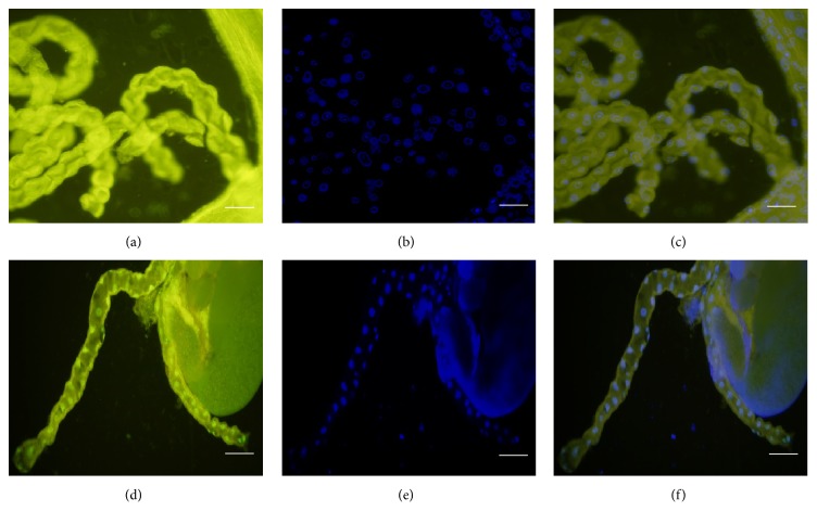
Fluorescence micrographs demonstrating TUNEL-positivity in Malpighian tubules. (a) TUNEL-staining of a Malpighian tubule of col4a1 +/− L3-larva incubated at 20°C; (b) DAPI-staining; (c) merge, tubules appearing TUNEL-negative. (d) TUNEL-positive Malpighian tubule of a col4a1 +/− L3-larva incubated at 29°C; (d) DAPI; (e) merge. Scale bars: (a)–(c) 50 μm, (d)–(f) 100 μm.

