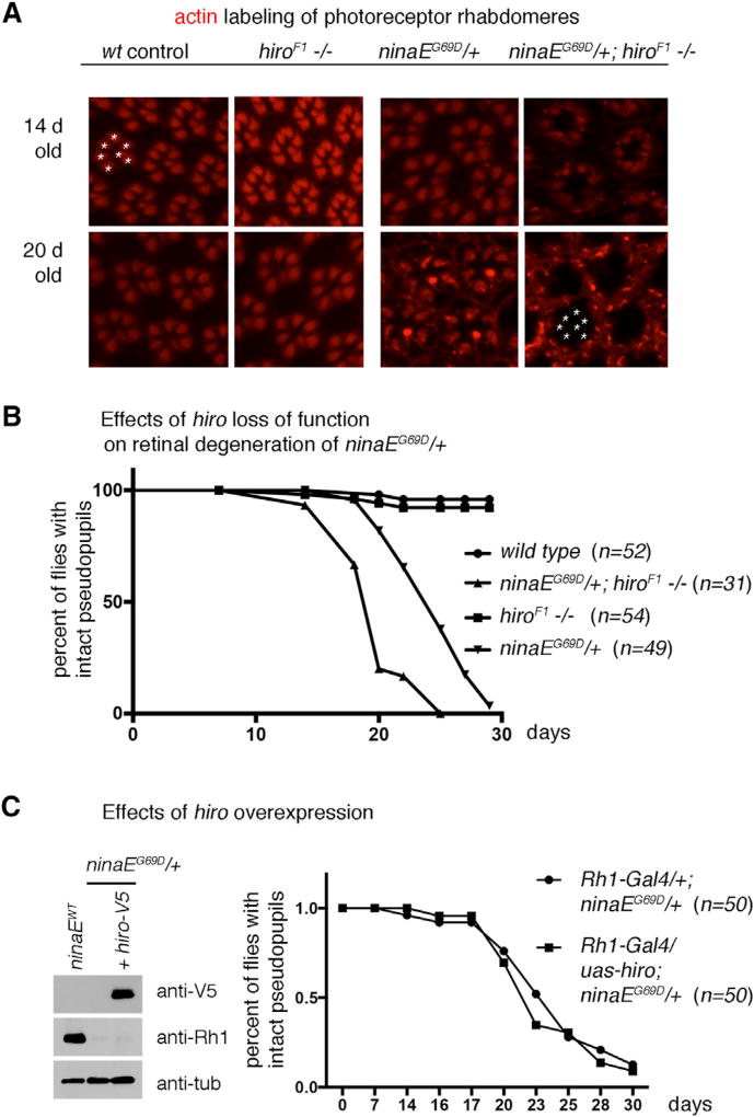
Figure 3. Loss of hiro Accelerates the Course of Age-Related Retinal Degeneration in ninaEG69D/+ Flies.
(A) Representative images of dissected ommatidia labeled with phalloidin that labels rhabdomeres of photoreceptors (red). The genotypes are indicated on top of each panel. Intact ommatidia show a trapezoidal arrangement of seven photoreceptor rhabdomeres (one example marked with asterisks on the upper left panel). Twenty-day-old ninaEG69D, hiroF1−/− eyes (lower rightmost panel) lack the trapezoidal pattern of phalloidin labeling. The asterisks in that panel indicate the expected positions of phalloidin-positive rhabdomeres in an ommatidium.
(B and C) Retinal degeneration was assessed in live flies containing Rh1-GFP to visualize photoreceptors, quantified, and exhibited as a graph.
(B) The effect of hiro loss of function on retinal degeneration. The genotype and the numbers (n) are indicated.
(C) The effect of hiro overexpression. UAS-hiro-V5 was expressed in ninaEG69D/+ photoreceptors using Rh1-Gal4. (Left) Western blot showing V5 epitope detection in the adult fly head extracts (top gel) and hiro-V5’s effect on Rh1 levels (middle gel). Anti-β-tubulin was used as a loading control. (Right) Comparison of the course of age-related retinal degeneration in control ninaEG69D/+ flies with those that express hiro-V5 in an otherwise identical genetic background.

