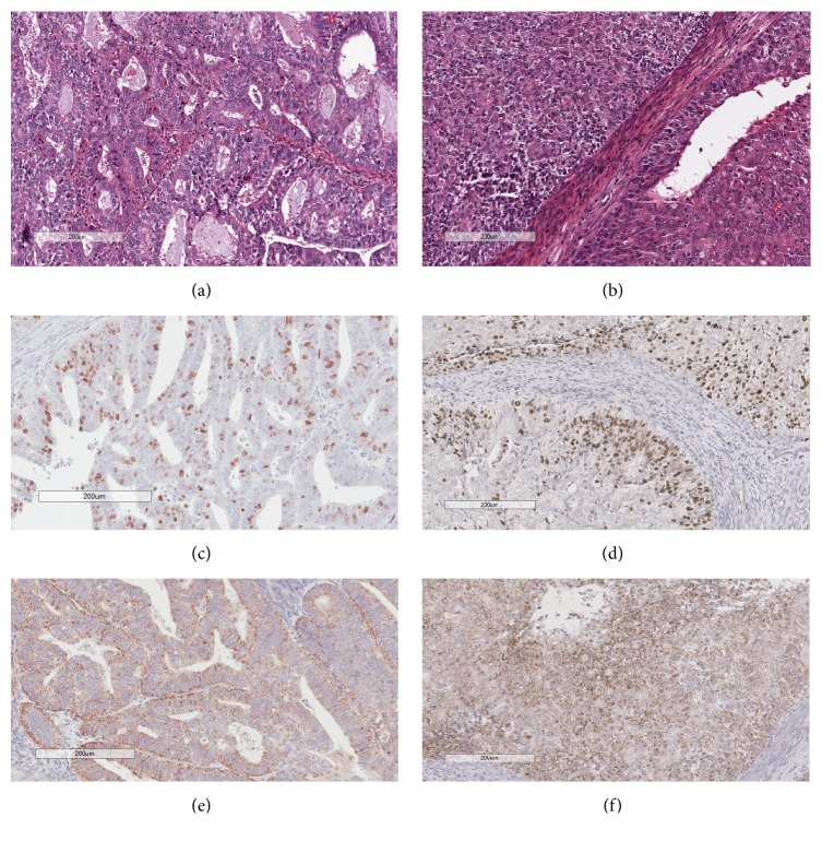
Figure 5.
Histopathological and immunohistochemical evaluation of endometrial carcinoma. (a) and (b) H&E stain of endometrioid endometrial carcinoma, histological grades I and II, respectively; (c) and (d) Ki67 stain on grade I and II sections, respectively; (e) and (f) IGSF9 stain on grade I and II sections, respectively. Both immunostains were quantified with the image analysis tools from Aperio digital pathology system. Numeric data were used in statistical analysis.

