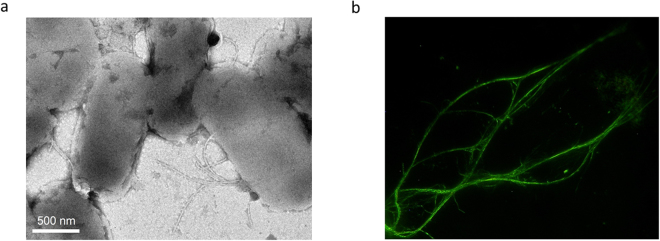
Figure 3.
Flagellar nanotube formation from FliC-aGFP_ENH_V2 subunits in vivo and in vitro. (a) Bright-field TEM image of Salmonella cells that possess mutant flagellar filaments. Samples were stained with 2% phosphotungstate to enhance image contrast. (b) Filaments polymerized from purified subunits as visualized by dark-field optical microscopy. Polymerization experiments were carried out in PBS buffer (pH 7.4) at 2 mg/ml protein concentration and 4 M AS was added to 0.6 M final concentration to initiate filament formation.

