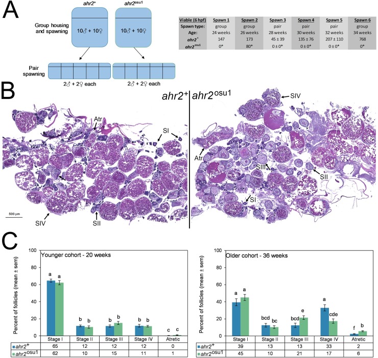
Fig 4. Reproductive impacts on the ovary are evident in ahr2osu1 zebrafish.
(A) A fecundity study was performed with group and pair spawning between weeks 24 and 34 with age-matched cohorts of ahr2+ and ahr2osu1 zebrafish. Weeks 24–34 correspond to the optimal reproductive period. Between spawns, pairs were re-combined into groups so that each pair spawn were randomly selected groups. *p < 0.05 for group spawning (Fisher’s exact test) or pair-wise spawning (Student’s t-test) when compared to wild type. For group spawns, n = 10 males, 10 females. For pair spawns, n = 5, each with 2 males and 2 females. (B) Ovarian histopathological assessments were performed on ahr2+ and ahr2osu1 zebrafish to quantify the representative follicle distribution. Photomicrographs for the older cohort (36 weeks) are shown for each genotype using the sections most representative of group averages. (C) Differential follicle analysis was performed for reproductively active adult zebrafish at 20 and 36 weeks (n = 4 per genotype for each age group). Statistical differences between genotypes and developmental stages were determined using two-way ANOVA with Tukey post hoc test for multiple comparisons (p < 0.05). Significance is indicated using compact letter display, and bars not in the same letter group are significantly different. Follicles were scored as Stage I (pre-vitellogenic primary growth), Stage II (early-vitellogenic cortical alveolus stage), Stage III (mid-vitellogenic), Stage IV (late-vitellogenic mature), and atretic.

