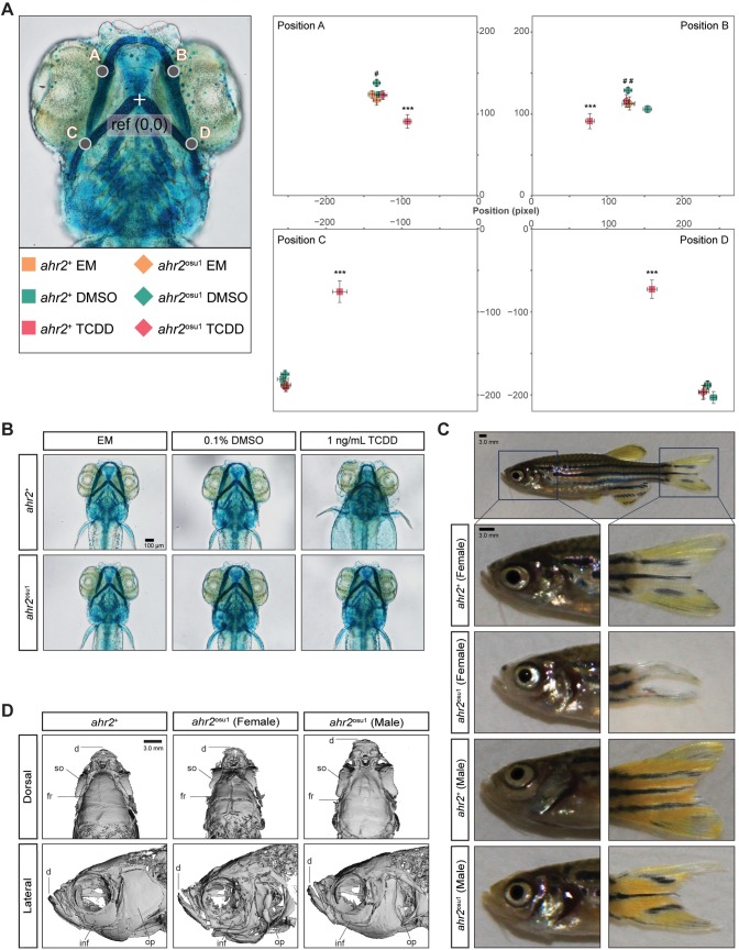
Fig 5. Fin and skeletal abnormalities observed in adult ahr2osu1 line.
Wild types and AHR2-null zebrafish were developmentally exposed at 6 hpf to EM, 0.1% DMSO, or 1 ng/mL TCDD, and the cartilage was stained and measured at 5 dpf. (A) A morphometric system was used to measure the position and length of landmark structures in the developing jaw. The position of jaw structures representing junctions between Meckel’s and palatoquadrate cartilages (points A and B) and the hyosymplectic and ceratohyal cartilages (points C and D) was measured relative to a reference point as shown. Statistical significance was determined by a modified two-way ANOVA with a Tukey post hoc test. Morphometric values represent mean ± SD (n = 9–10; p < 0.05 = * or #, p < 0.01 = ** or ##, p < 0.001 = ***). The asterisk (*) indicates statistical significance in wild-type fish exposed to TCDD compared to wild-type fish exposed to DMSO, and (#) indicates statistical significance in DMSO-exposed ahr2osu1 mutants compared to DMSO-exposed wild-type fish. (B) Representative ventral views of 5 dpf cartilage in wild types and AHR2-null mutants developmentally exposed to EM, 0.1% DMSO, or 1 ng/mL TCDD. Black bar in bottom right corner = 100 μm. (C) Representative brightfield images of adult wild types’ and ahr2osu1 mutants’ heads and caudal fins. Dark blue square represents area in panels below. Black bar in top left corner = 3.0 mm. (D) microCT imaging of adult wild-type (female) and ahr2osu1 mutant female and male zebrafish heads. Male and female wild-type microCT scans looked the same and a representative female image was selected as the representative image. Notable differences were observed in the dentate (d), supraorbital (so), frontal (fr), infraorbital (inf), and operculum (op). Black bar in top right corner = 3.0 mm.

