
Fig 1. Optical coherence tomography imaging protocol and analysis, for both foveal-centered and temporally centered scans.
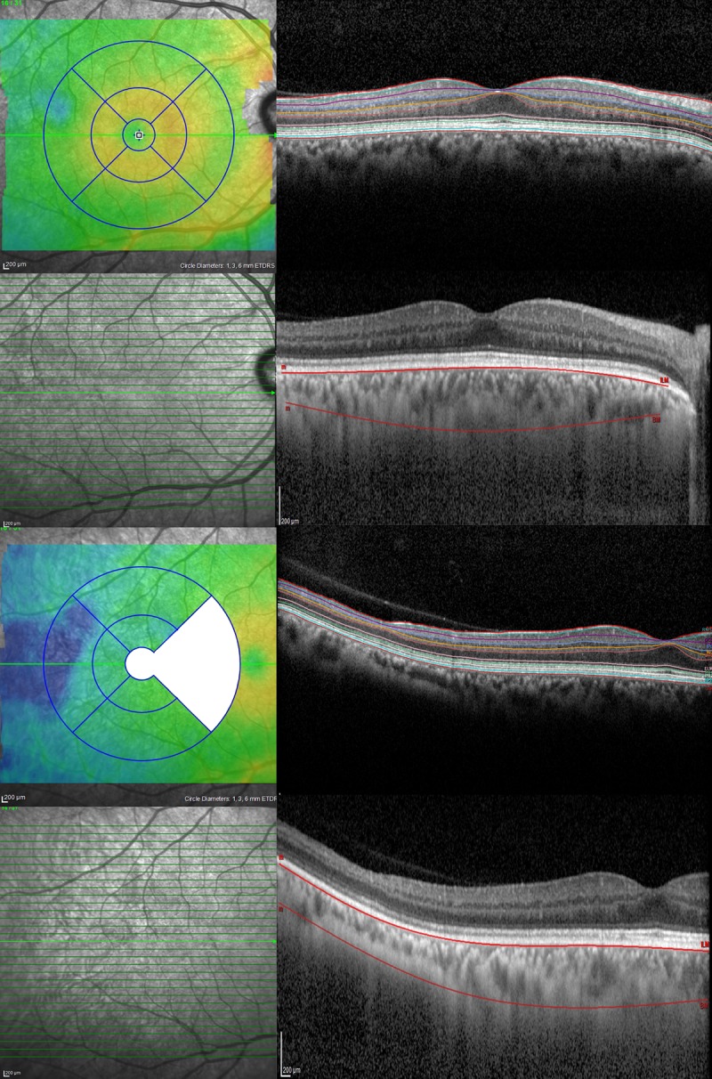
(Left column) Color-coded retinal thickness maps were generated by the built-in software of the device, which then automatically applied the ETDRS grid after scanning eyes using 31 high-resolution B-scans. The nasal quadrant and the central ring of the temporally-centered scan were systematically not measured because overlapping with the temporal quadrant of the foveal-centered scan. (Right column) The segmentation software of the SD-OCT device automatically detected each retinal layer, including retinal nerve fiber layer, ganglion cell layer, inner plexiform layer, inner nuclear layer, outer plexiform layer, outer nuclear layer, photoreceptor layer, and retinal pigment epithelium. Segmentation of the choroid was performed manually.