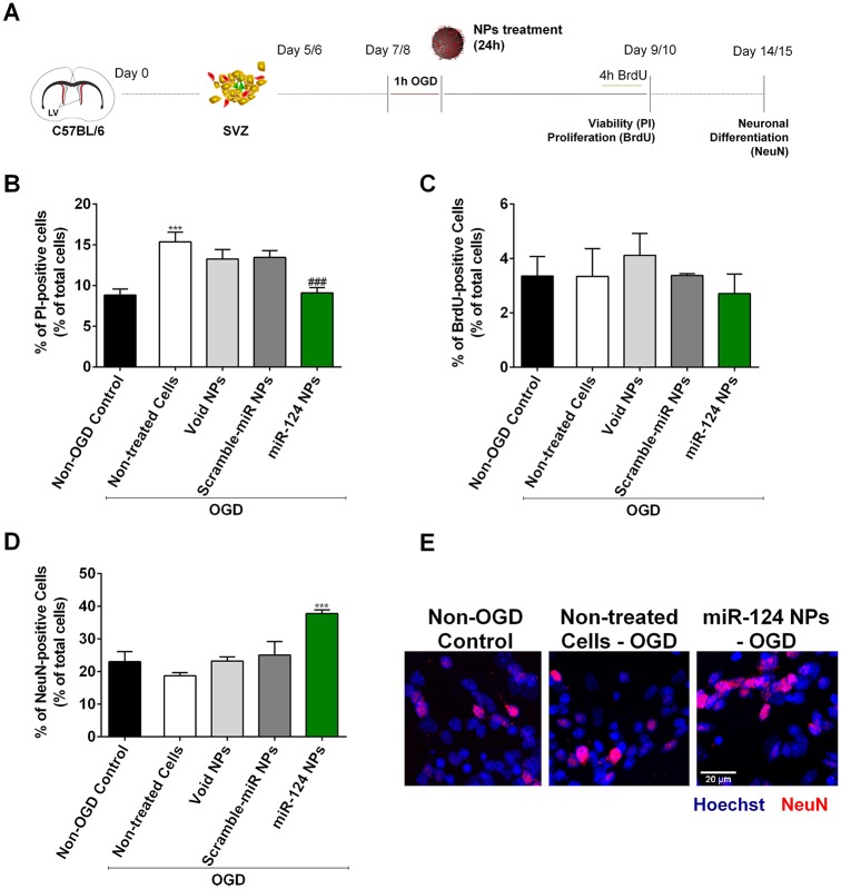
Fig 1. Effect of miR-124 NPs treatment on SVZ cell cultures after OGD.
(A) Experimental design of in vitro experiments. NSCs where isolated from the SVZ of C57BL/6 J 1 to 3 days-old pups and grown in suspension for 5 or 6 days to obtain neurospheres. Neurospheres were seeded and allowed to grow as monolayer for 2 days before being stimulated with OGD for 1 h. Cells were then incubated with void NPs, scramble-miR NPs or miR-124 NPs for 24 h. Cells were maintained in culture according to the parameters evaluated: 48 h for cell viability and proliferation assays and 7 days for neuronal differentiation. (B) Cell viability assessed by incorporation of propidium iodide (PI) into dead cells and presented as percentage of PI-positive cells in cultures stimulated with OGD and either non-treated or treated with void NPs, miR-scramble NPs or miR-124 NPs, respectively. PI-positive cells quantified in normoxic cultures (non-OGD control) served as controls. (C) Proliferation of SVZ cultures after OGD followed by different treatments. Graphs show the percentage of BrdU-positive cells of total cell counts. D) Neuronal differentiation of the cultures measured by the percentage of NeuN-positive cells in NSC monolayer cultures. (E) Representative fluorescence photomicrographs of NeuN immunostainings in non-OGD control cultures, OGD non-treated and OGD miR-124 NPs treated cultures 7 days after treatment. Nuclei are shown in blue and NeuN in red. Scale bar: 20 μm. Data are expressed as means ± SEM (n = 3). Statistical analysis was performed using one-way ANOVA and Tukey multiple comparison. ***p < 0.001 versus non-OGD control; ### p < 0.001 versus 1h OGD non-treated cell condition. Abbreviations: NeuN, neuronal nuclei; NPs, nanoparticles; NSCs, neural stem cells; OGD, oxygen and glucose deprivation; SVZ, subventricular zone.

