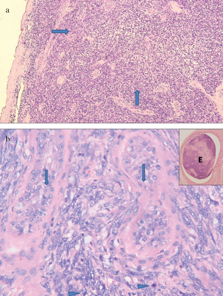
Figure 2. a, b.

Microscopic examination of tumor. (a) Oval to spindle- shaped tumor cells arranged in interlacing bundles and fascicles (arrows) (H&E, 100X). (b) Tumor cells showing moderate nuclear atypia and frequent mitoses (arrowhead) with entrapped seminiferous tubules (arrows) (H&E, 400X). Inset shows vascular tumor embolus (E)
