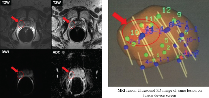
Figure 2.
The different mpMRI sequence images and 3D images of prostatic lesion of a 69-year-old active surveillance patient from our clinic. His last PSA was 4.3 ng/mL, and has Gleason grade 3+3 PCa. mpMRI was performed by 1.5T magnet device with ERC.
- Prostate volume is estimated 19 cc.
- Dominant lesion measures 1.1 × 1.0 cm and is located in the right peripheral zone at the level of the midgland at 7–9 o’clock position.
- Moderate low T2 signal, ADC signal is reduced.
- Suspicious focal enhancement is seen. Qualitative suspicion of clinically significant disease: 4. Likely.
- There is indistinctness between the lesion and the prostatic capsule on the coronal images, suspicious for early extraprostatic extension.

