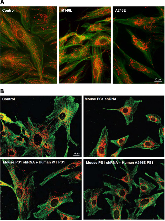
Fig. 1.
a Representative mitochondrial morphology in AD-mutant cells. Human fibroblasts were stained with MitoTracker (red) and anti-tubulin (green). Note the relatively dispersed distribution of the mitochondria in the control, whereas they are more perinuclear in the FAD-PS1M146L and FAD-PS1A246E cells. b MEFs in which PS1 was knocked down (by small hairpin RNA). Cells were stained as in a. Note relatively dispersed distribution of the mitochondria in the control, whereas they are more perinuclear in the PS1-knockdown cells. This phenotype could be rescued by overexpression of WT human PS1, but not by expression of a human pathogenic PS1 mutation (A246E)

