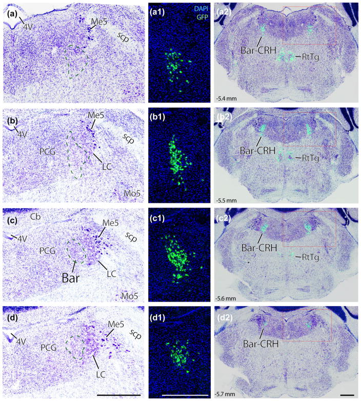
FIGURE 1.
Nissl cytoarchitecture in the mouse brainstem region that contains Barrington’s nucleus (Bar). Panels (a–d): successive rostral-to-caudal sections through the pons of a Crh reporter mouse. Every third section (35 μm-thick) is shown across a total span of ~0.5 mm. Bar neurons, using Nissl cytoarchitecture alone, are evident as an ovoid cluster of medium-large neurons located medial to the ventral locus coeruleus (dashed line in panel c). Rostral and caudal to this core, we cannot distinguish Bar neurons from surrounding populations in a Nissl preparation. Panels (a1–d1) show a GFP reporter for Crh in the same tissue sections as (a–d), at the same magnification, in fluorescence images taken prior to Nissl counterstaining. The location of BarCRH neurons in each section is indicated by a green dashed line in panels (a–d). Panels (a2–d2) show the distribution of BarCRH neurons throughout each section by blending images of GFP and thionin counterstaining from the same sections. Scale bars are all 500 μm (d–d2), and apply to the column above. Approximate level in mm caudal to bregma is shown in the lower left corner of panels (a2–d2) and applies to all panels in each row. 4V = fourth ventricle; Bar-CRH = corticotropin releasing hormone neurons in Barrington’s nucleus; LC = locus coeruleus; Me5 = mesencephalic nucleus and tract of the trigeminal nerve; Mo5 = motor nucleus of the trigeminal nerve. PCG = pontine central gray matter; RtTg = reticular tegmental nucleus; scp, superior cerebellar peduncle

