FIGURE 10.
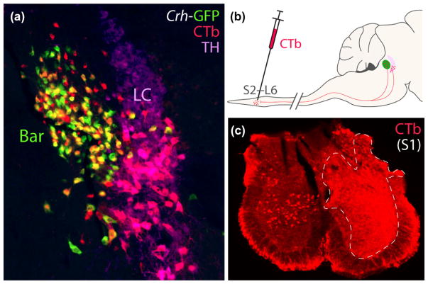
Injecting CTb into the lumbosacral spinal cord retrogradely labels neurons in Bar and LC. (a) In this CRH reporter mouse (Crh-IRES-Cre; L10-GFP), a slight majority of retrogradely labeled neurons in Bar express GFP. These CTb-labeled, BarCRH neurons appear yellow-orange due to labeling for both CTb (red) and GFP (green). Outside Bar, CTb-labeled neurons were located in the ventral LC and subcoeruleus, and all of them express TH. CTb-labeled LC neurons appear pink due to labeling for both CTb (red) and TH (magenta). The diagram in panel (b) shows the CTb injection located in the lumbosacral spinal cord. Panel (c) shows a level of the sacral spinal cord in this mouse at the center of the injection site resulting in the ipsilateral, retrograde labeling shown in (a); the contralateral sacral gray matter contains several CTb retrogradely labeled neurons. Scale bars are 200 μm

