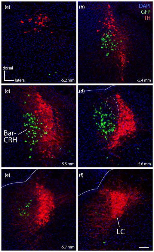
FIGURE 2.
The crescent-shaped locus coeruleus (LC) cradles the lateral surface of mouse BarCRH neurons. Panels (a–f) show the close relationship between LC (immunofluorescence labeling for tyrosine hydroxylase; TH, red) and BarCRH (GFP reporter for Crh, green). BarCRH neurons are found at middle-to-rostral levels of the LC (panels B–E, corresponding to the same approximate bregma levels as Figure 1, panels a–d). No neurons are double-labeled. Rostral to the LC, there are no more BarCRH neurons, though sparse, large neurons immunoreactive for TH extend rostrally into the periaqueductal gray. At caudal levels (panels e, f), there are few or no BarCRH neurons. At these caudal levels, LC dendrites project medially into a zone immediately behind the core of Bar. At central levels containing the core of Bar (panel d), its dorsolateral surface diverges from the dorsal LC; the resulting gap between BarCRH and the dorsal LC (dashed outline in panel d) is filled by a wedge of FoxP2/glutamatergic neurons, which are shown in Figure 6. Lines in panels (d–f) mark the ependymal surface of the fourth ventricle. Scale bar is 100 μm and applies to all panels

