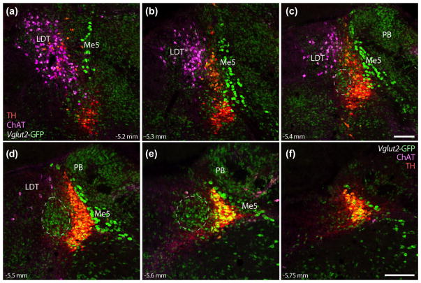
FIGURE 5.
Glutamatergic neurons in Bar are surrounded by mutually exclusive populations of cholinergic and catecholaminergic neurons, as well as other glutamatergic populations. Panels (a–f) show, in successive rostral to caudal levels, the relative positions of neurons in each population. Laterodorsal tegmental nucleus (LDT) neurons, labeled by immunofluorescence for choline acetyltransferase (ChAT, magenta), are located rostral and medial-dorsal to Bar. Vglut2 GFP reporter is found in many LC catecholaminergic (red +green = yellow) and LDT cholinergic neurons (magenta +green = white), as well the large Me5 neurons dorsolateral to the LC, and most neurons in the PB. The dashes in panels (c–e) outline the approximate location of Bar, estimated from the distribution of BarCRH neurons shown above. Scale bars are all 200 μm (panel c applies to a–c; panel f applies to d–f). Approximate levels in mm caudal to bregma are given at the bottom left or right of each panel

