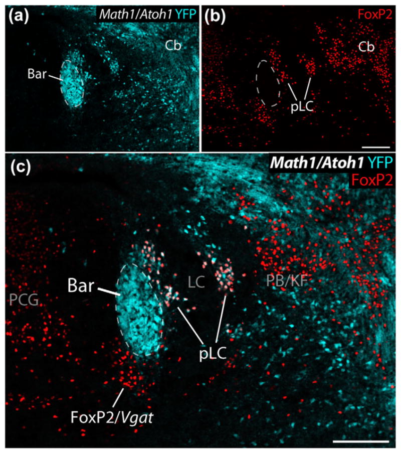
FIGURE 7.

In this Cre-reporter mouse (Atoh1-Cre; R26-lsl-YFP), YFP expression (ice-blue) reveals cells derived from embryonic Math1/Atoh1-expressing precursors in the rhombic lip neurothelium in a P0 mouse. Among the Math1/Atoh1-derived neurons in the pontine tegmentum are a dense cluster with the expected location, size, and shape of Bar. These images also contain FoxP2 immunolabeling (red) in cells surrounding Bar. LC neurons, which are derived from separate precursors, do not express YFP. Cerebellar cells (granule cell precursors) and other neurons express YFP, including FoxP2 (pLC) neurons bordering the dorsolateral surface of Bar and winding through the LC. In contrast, FoxP2 neurons in the PCG, including those bordering the ventromedial surface of Bar (FoxP2/Vgat), do not express YFP. Other populations highlighted in this image include a group of non-Math1/Atoh1-derived, FoxP2+ neurons in the caudal PB/Kolliker-Fuse nucleus (PB/KF), which are GABAergic and are separate from the larger population of Math1-derived (glutamatergic) FoxP2 expressing neurons through much of the PB found at levels rostral to this. Scale bars in panels (b) and (c) are 200 μm
