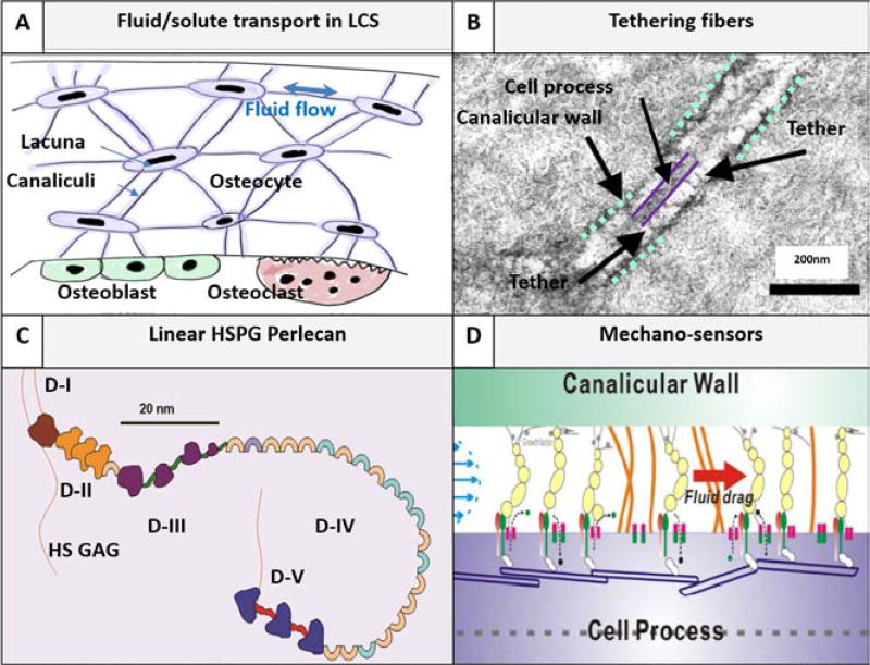
Fig. 1.
Pericellular matrix (PCM)-filled lacunar-canalicular system (LCS) is bone’s natural chromatographic column, where fluid/solute transport to and from osteocytes is regulated. (A) Fluid and solute transport in the LCS is critical for osteocyte survival (e.g., nutrient supply and cell-cell signaling) and proper function (e.g., mechanosensing) [7]. (B) The canalicular channel contains an electron-dense PCM and transverse fibers tether the cell process with the canalicular wall as seen under transmission electron microscopy [47]. (C) Schematic of perlecan/HSPG2, a proteoglycan, consisting of five domains and four HS sidechains (Courtesy of M.C. Farach-Carson). Perlecan is the first identified PCM component [48] with a contour length of 170 nm and diameter of ~4 nm [51]. (D) Perlecan as well as other tethering fibers regulate the fluid and solute transport in the LCS. The PCM fiber density (inversely related to fiber spacing) determines the LCS’s hydraulic permeability, the magnitude of fluid velocity and the fluid drag force so that osteocytes can sense and respond to mechanical loading [43]. The PCM fiber density also modulates solute diffusion and convection due to steric exclusion between finite sized solutes and the PCM fibers [35]. The osteocyte PCM fiber spacing is found to be 10.3, 13.4, 17.4 nm in young adult, aged, and aged perlecan deficient LCS, respectively [35].

