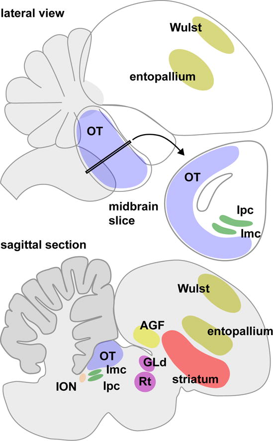
Figure 3. Schematic diagram of the bird brain.

The lateral view illustrates the prominent position of the optic tectum (OT) in the midbrain, and outlines the locations of the visual Wulst and entopallium in the forebrain. The midbrain slice shows the spatial relationship between the optic tectum, and the adjacent nucleus isthmi pars parvocellularis (Ipc) and nucleus isthmi pars magnocellularis (Imc). The sagittal section provides a cut-away view that makes other attention-related structures visible: the isthmo-optic nucleus (ION) in the midbrain, the nucleus rotundus (Rt) and lateral geniculate nucleus pars dorsalis (GLd) in the thalamus, and the striatum and arcopallial gaze field (AGF) in the forebrain.
