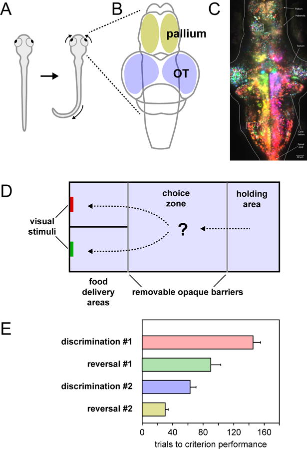
Figure 5. Brain and behavior in the zebrafish.

A. Dorsal view of a zebrafish illustrating the J-turn tail movement and convergent eye movements elicited by the presentation of a visual prey stimulus. B. Dorsal schematic view of the zebrafish brain showing the locations of the optic tectum and pallium. C. Image of larval zebrafish brain obtained using light-sheet microscopy while animal was presented with moving visual stimuli. Because the brain is small and the tissues are transparent, it is possible to image individual neurons throughout the tectum and pallium. Individual neurons are color-coded based on their preferred direction of motion, with magenta indicating preference for rightward motion and yellow indicating preference for leftward motion. Image reproduced with permission from (Freeman et al., 2014). D. Schematic diagram of the aquarium tank used in a 2-alternative choice task. After being introduced in the holding area, a trial was started when the opaque barriers were removed and the fish gained access to the choice zone and food delivery areas. Fish expressed their choice by swimming into one of the two food delivery areas and approaching the colored visual stimulus. E. The number of trials required to reach criterion performance (6 consecutive correct trials) during the four phases of the experiment. Panels (D) and (E) adapted with permission from (Parker et al., 2012).
