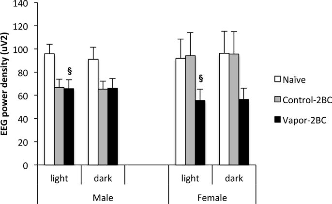
Figure 8.

Delta (0.5-4.0 Hz) power values obtained during SWS across 12 hour light and dark periods are shown. Values (Mean +/− SEM) were compared using a two way ANOVA and Fisher’s PLSD tests. Vapor-2BC mice had decreased delta power relative to naïve mice during the light phase. § p < 0.05 vs. naïve, sexes combined.
