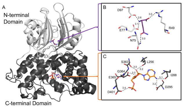
Fig. 1.
Model of human ASNS protein structure. A human ASNS protein model was generated utilizing the crystal structure of E. coli asparagine synthetase B (PDB: 1CT9) as a template and then adapted to contain variants observed in patients with ASD, as described in the Methods section. (Panel A) N-terminal (light grey) and C-terminal (dark grey) domains with the substrate glutamine (purple) and the product AMP (orange) shown as sticks. (Panel B) Glutamine binding pocket illustrating glutamine hydrogen bonds. (Panel C) ATP/AMP binding pocket with AMP binding shown. Hydrogen bonds are represented as black dashes and the distance of each is shown in angstroms (Å).

