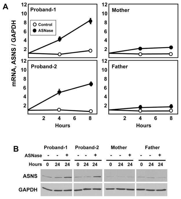
Fig. 5.
Limiting extracellular asparagine causes up-regulation of the ASNS gene in the proband cells. Skin fibroblasts from the parents and both probands were maintained in culture and then incubated in control DMEM medium or DMEM containing 0.1 U/ml of ASNase to deplete extracellular asparagine. After incubation for the indicated period of time, total RNA and protein was isolated. Real-time quantitative PCR was performed to measure steady state mRNA levels for ASNS and GAPDH (Panel A). The data are the averages ± standard deviations of three determinations within an experiment. Each experiment was repeated at least once to verify qualitative reproducibility. Where not visible, the standard deviation bars are contained within the symbol. For protein analysis, immunoblots were performed using antibodies specific for ASNS and GAPDH, as described in the Methods section (Panel B). Each sample was tested at least twice to assess reproducibility of the results and a representative blot is shown.

