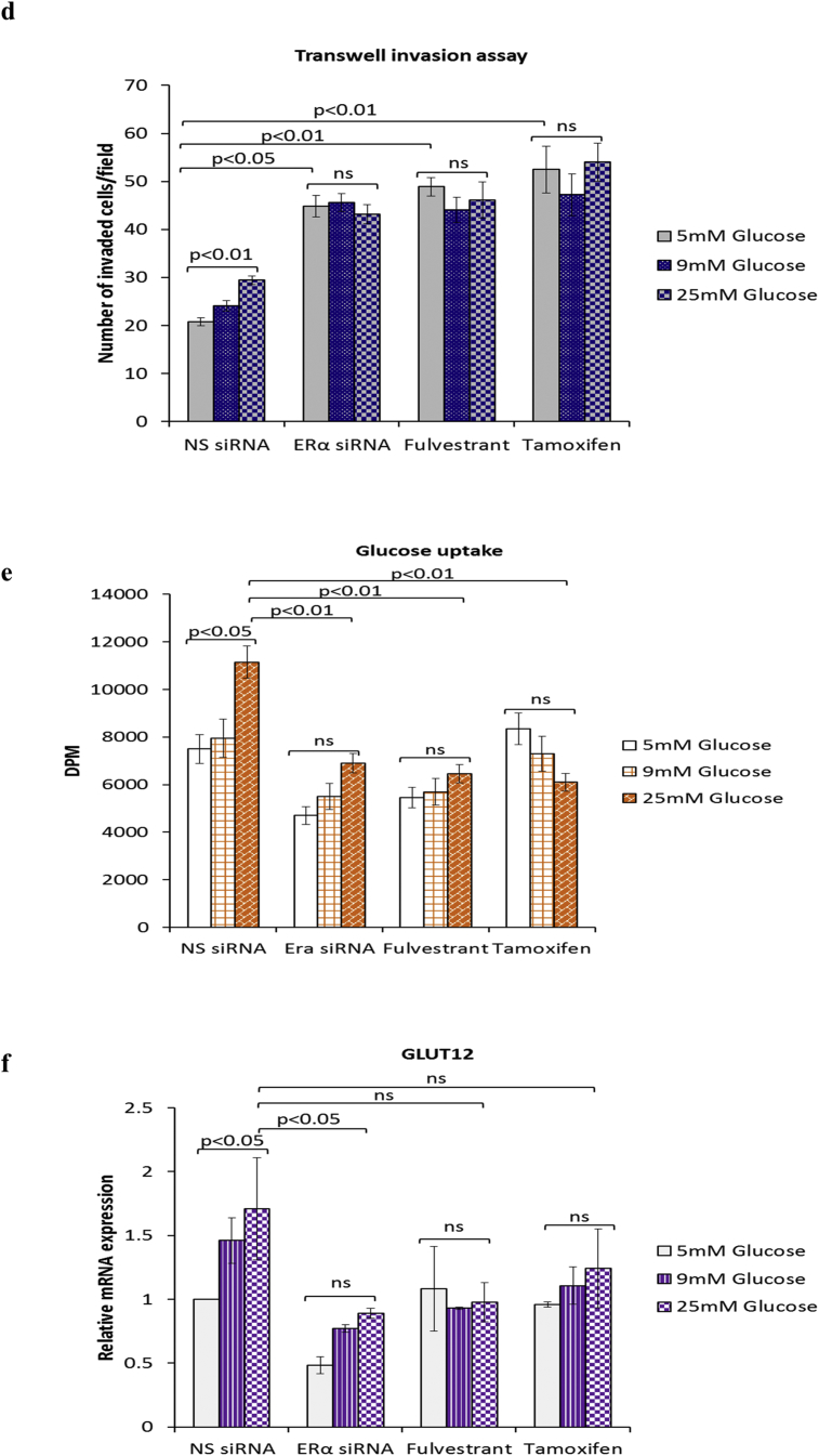
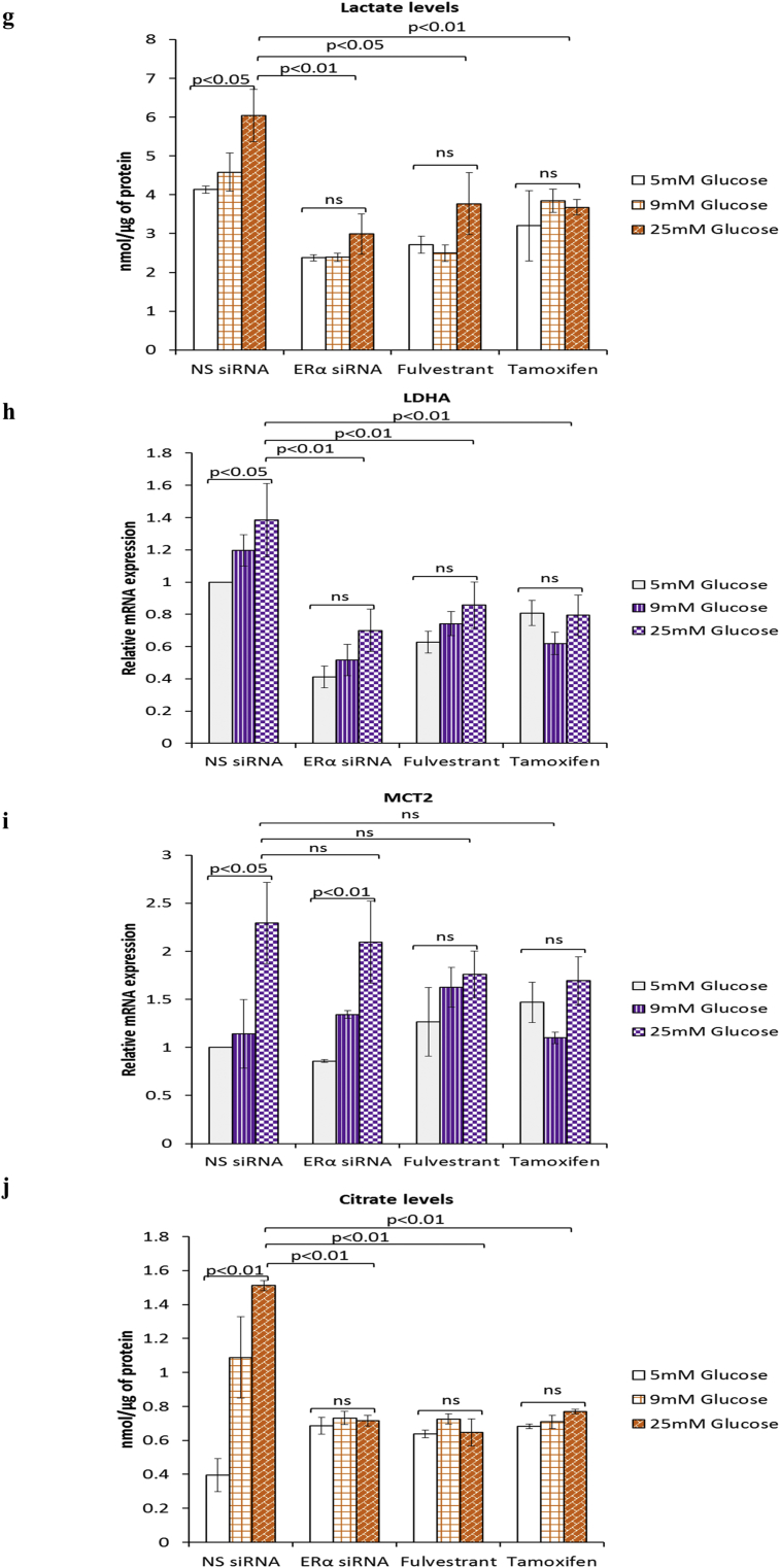
Fig. 4.
Inhibition of ERα during hyperglycaemia-induced, matrix-specific EMT results in a more metastatic phenotype irrespective of the glucose concentration. (a, i) Protein expression of EMT markers following downregulation of ERα signalling by siRNA or anti-estrogens fulvestrant (0.1 μM) and tamoxifen (1 μM). (a, ii) The densitometry measurements from the western blot are shown. (b) Quantification of SLUG mRNA levels as determined by qPCR. (c, upper panel) Cell growth was assessed as described above. (c, lower panel) Western blot detection of PARP cleavage was used as an indicator of apoptosis. (d) Cell invasion was measured as described above. (e–i) Changes to the metabolic parameters following ERα knock down by siRNA and treatment with antiestrogens were assessed as described in the legend of Fig. 3. Results shown are representative of three independent experiments, each performed in triplicate. Data are represented as mean ± SEM. (NS = non-silencing).


