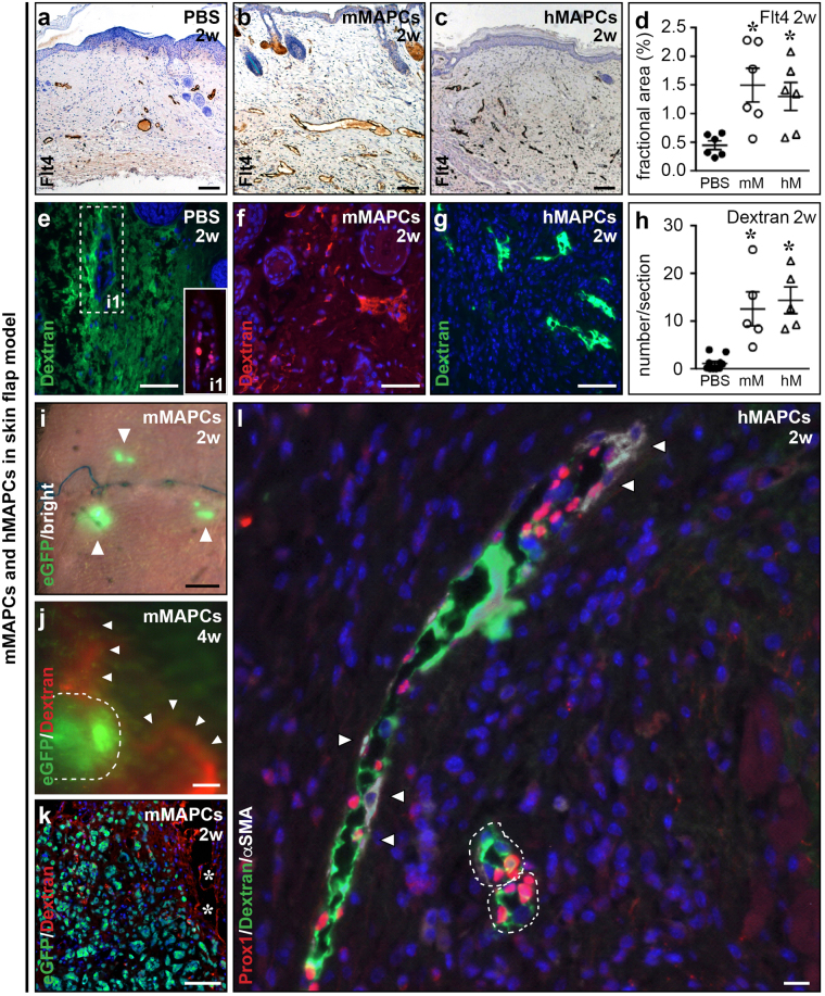
Figure 4.
MAPCs restore lymphatic capillaries and pre-collectors. (a–d) Flt4-stained wound cross-sections from PBS (a), mMAPC (‘mM’; b) or hMAPC-treated (‘hM’; c) mice, and corresponding quantification (d; n = 6; P = 0.0074 by Kruskal-Wallis test; *P < 0.05 versus PBS by Dunn’s post-hoc test). (e–h) Wound cross-sections from PBS (e), mMAPC (f) or hMAPC-treated (g) mice revealing functional (dextran (red or green)-perfused) lymphatics in cell-treated mice, and corresponding quantification (h; n = 5–10; P < 0.0001 by Kruskal-Wallis test; *P < 0.05 versus PBS by Dunn’s post-hoc test). Inset (i1) in e shows corresponding Prox1-stained (in red) region. Note diffuse fluorescence signal in e representing FITC-dextran that failed to be drained. (i) Merged bright field/fluorescence image of the wound transplanted with eGFP+ mMAPCs (in green; indicated by arrowheads) 2 w earlier. (j) Merged green/red fluorescence images of the wound transplanted with eGFP+ mMAPCs (circled by dashed line) 4 w earlier. Note Rhodamin-dextran-filled lymphatic vessels (red; indicated by arrowheads) in the vicinity of transplanted cells. (k) Cross-section through the wound, revealing transplanted eGFP+ mMAPCs (in green) adjacent to functional (red Rhodamin-dextran-filled) lymphatics (asterisks). (l) Merged picture of green (FITC-labelled dextran), red (Prox1) and far-red (αSMA) fluorescence images of a wound transplanted with hMAPCs 2 w earlier, revealing a functional sparsely αSMA-coated (indicated by arrowheads) Prox1+ lymphatic pre-collector and two functional Prox1+/αSMA− lymphatic capillaries (circled by white dashed lines). Haematoxylin or DAPI were used to reveal nuclei in a–c and e–g, k, l, respectively.

