Fig. 1.
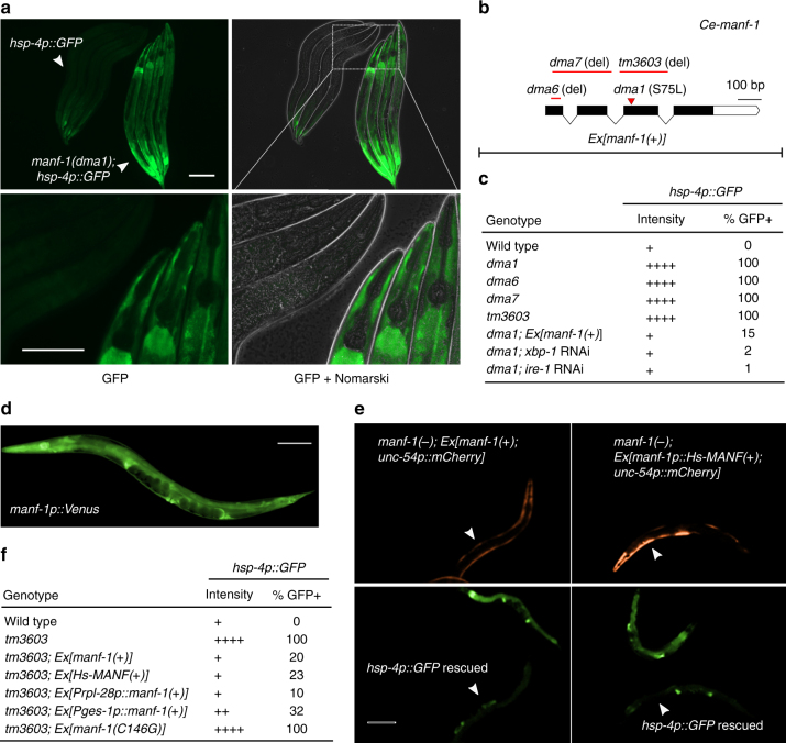
A genetic screen identifies C. elegans manf-1 in regulating hsp-4p::GFP. a Exemplar GFP fluorescence and Nomarski images showing constitutive activation of hsp-4p::GFP in the dma1 mutant isolated from mutagenesis screens. Wild type and mutant L4-stage animals (indicated by arrows) are aligned in the same field to compare GFP intensity at low (above) or high (bottom) magnification. Scale bars, 100 µM. b Schematic of manf-1 gene structure with various manf-1 alleles and extrachromosomal arrays (Ex) indicated. c Summary table for the hsp-4p::GFP phenotype of the wild type compared with various mutants, with indicated levels of GFP intensity in the intestine, the major site of hsp-4p::GFP expression, and penetrance (N ≥ 100 for each genotype). d Integrated manf-1p::Venus transcriptional reporter indicating the widespread expression of manf-1. e Exemplar GFP fluorescence images showing the rescue of the hsp-4p::GFP phenotype in the manf-1 null tm3603 mutant by C. elegans manf-1(+) and human MANF driven by the manf-1 promoter. Transgenic rescuing arrays are marked by unc-54p::mCherry expressed in body wall muscles. f Summary table showing the rescuing effect of various transgenes on hsp-4p::GFP of the manf-1(tm3603) mutant with indicated GFP intensity levels and penetrance (N ≥ 100 for each genotype)

