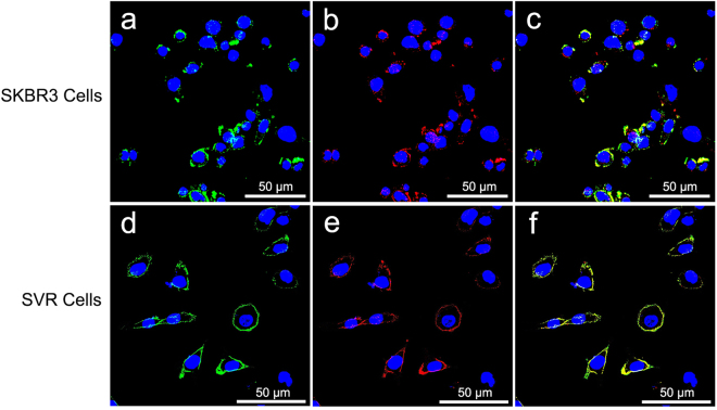
Figure 3.
Confocal microscope images of SKBR3 and SVR cells incubated with dual-targeted NBD. (a–c) SKBR3 cells were substantively stained by nanoparticle-antibody bioconjugates (green, red or yellow, incomplete circles) around cell nuclei. (d–f) SVR cells were strongly stained by nanoparticle-antibody bioconjugates (bright, complete green, red or yellow circles). Green, fluorescence signal of anti-HER2 antibody labeled with FITC. Red, fluorescence signal of anti-VEGFR2 antibody labeled with PE. Yellow, merged fluorescence of red and green. Blue, nuclei stained with DAPI.

