Figure 5.
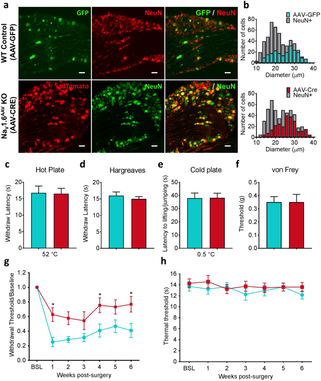
AAV-Cre mediated NaV1.6 knockout in adult mice attenuates SNI-induced mechanical allodynia. (a) Representative images of L4 DRG sections showing AAV-GFP and AAV-Cre infected DRG neurons, which are marked by GFP and tdTomato, respectively. Neurons were identified by the neuronal marker NeuN. Scale bar = 50 µm. (b) Size distribution of AAV-GFP and AAV-Cre infected DRG neurons. (AAV-GFP: expressed in 245 out of 485 L4 DRG neurons, n = 3; AAV-Cre: expressed in 275 out of 502 L4 DRG neurons, n = 4). (c) Noxious heat thresholds measured by hot plate test in AAV-GFP control (WT control, n = 8) and AAV-Cre mediated NaV1.6 knockout (NaV1.6AAV KO, n = 15). (d) Noxious heat thresholds: Hargreaves test (WT control, n = 7; NaV1.6AAV KO, n = 11). (e) Noxious cold thresholds: cold plate test (WT control, n = 5; NaV1.6AAV KO, n = 7). (f) Noxious mechanical thresholds measured using von Frey test (WT control, n = 6; NaV1.6AAV KO, n = 11). (g) Mechanical allodynia following SNI was measured by the von Frey test in WT control (n = 10) and Nav1.6AAV KO mice (n = 12). Difference between the two groups was significant at week 1 (p = 0.0226), week 4 (p = 0.0455) and week 6 (p = 0.0336) post-surgery. (h) Thermal threshold measured by Hargreaves test was not changed compared to baseline in both WT control (n = 14) and Nav1.6AAV KO mice (n = 15). Data are presented as mean ± SEM. *p < 0.05 based on two-way ANOVA followed by Bonferroni post hoc test.

