Figure 6.
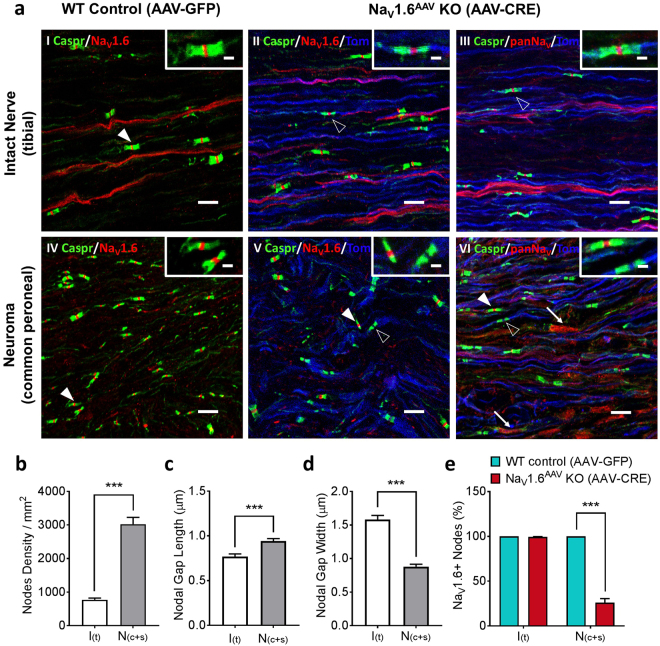
NaV1.6 knockout leads to empty nodes in myelinated fibers within the neuroma. (a) Confocal images showing NaV1.6 or panNaV (red) and contactin-associated protein (caspr, green) immunostaining as well as tdTomato signal (blue) in intact Tibial nerve (upper row) or neuroma (Common Peroneal nerve, bottom row) from AAV-GFP infected (WT control) and AAV-Cre infected (NaV1.6AAV KO) mice. Samples were collected 6 weeks after SNI and AAV injection. In intact nerve (aI) and neuroma (aIV) from wild-type mice, NaV1.6 protein is present in every node of Ranvier (filled arrowhead). In intact nerve from NaV1.6AAV KO mice NaV1.6 (aII) protein is stable in nodes (unfilled arrow head) along tdTomato positive fibers (blue, reporter for NaV1.6 gene excision); similar nodal staining along tdTomato positive fibers is observed when a pan sodium channel antibody is used (aIII, unfilled arrow head). In neuroma from NaV1.6AAV KO mice (aV), NaV1.6 protein is only present at nodes (filled arrow head) along tdTomato-negative fibers but absent at nodes along tdTomato-positive (blue) fibers (unfilled arrow head). Using a pan sodium channel antibody shows that the absence of Nav1.6 protein at nodes (unfilled arrow head) along tdTomato-positive (blue) fibers is not compensated by other NaV isoforms (aVI). Filled arrow heads point to representative nodes along tdTomato-negative A-fibers and unfilled arrow heads point to representative nodes along tdTomato-positive A-fibers. Arrows point to representative non-myelinated regions within the neuroma where sodium channels accumulate following injury. Scale bar = 10 µm; Inset scale bar = 2 µm. (b) Comparison of average density of NaV1.6-positive nodes in intact nerve (white, n = 10) and neuroma (grey, n = 10). (c) Comparison of average nodal gap length in intact nerve (white, n = 10) and neuroma (grey, n = 10). (d) Comparison of average nodal gap width in intact nerve (white, n = 10) and neuroma (grey, n = 10). (e) Percentage of NaV1.6-positive neurons in neuroma after AAV-Cre (NaV1.6AAV KO, n = 5) treatment or AAV-GFP (WT control, n = 5) treatment. Data are presented as mean ± SEM. ***p < 0.001 based on unpaired t test.

