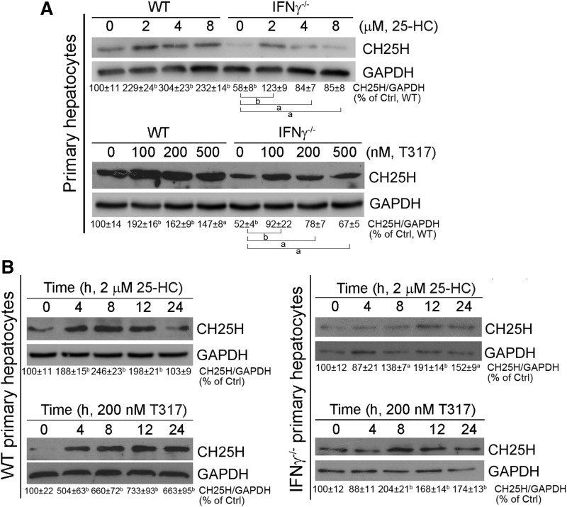
Fig. 6.
Lack of IFN-γ expression reduces but does not abolish LXR-activated hepatic CH25H protein expression. Primary hepatocytes were isolated from wild-type and IFN-γ−/− mice. A: comparison of CH25H protein basal levels or induction of CH25H protein expression by 25-HC or T317 treatment between wild-type and IFN-γ−/− hepatocytes. B: Wild-type and IFN-γ−/− hepatocytes were treated with 25-HC (2 μM) and T317 (200 nM) for the indicated times. Expression of CH25H protein was determined by Western blot. aP < 0.05, bP < 0.01 vs. control or as indicated (n = 3).

