Fig. 4.
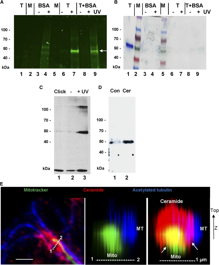
Cross-linking of pacFACer to tubulin and binding to ceramide. A, B: Tubulin from pig brain was cross-linked to pacFACer, tagged with Cy7.5 azide, and then separated by SDS-gel electrophoresis [fluorescence scanning (A); Coomassie staining (B)]. Lane 1, control tubulin (1 μg); lane 2, protein standard; lane 3, BSA − UV; lane 4, BSA + UV; lane 5, protein standard; 6, tubulin − UV; lane 7, tubulin + UV; lane 8, tubulin + BSA − UV; lane 9, tubulin + BSA + UV. The arrow points at the tubulin band. C: Protein from astrocytes was cross-linked to pacFACer, tagged with biotin azide, and the protein pulled down with streptavidin agarose beads. Immunoblot was performed using β-tubulin mouse IgG Lane 1, click reaction without pacFACer; lane 2, with pacFACer − UV; lane 3, with pacFACer + UV. D: Pull-down of protein with control agarose (Con) or ceramide-linked agarose beads (Cer). The immunoblot of protein in the eluate was probed with anti-detyrosinated α-tubulin rabbit IgG. E: High-resolution confocal microscopy showing arrangement of microtubules (MT), mitochondria (Mito), and ceramide (middle and right panels show z-stack reconstruction along dashed line in the left panel). The arrows point at contact structures of ceramide (CEMAM) with microtubules and mitochondria.

