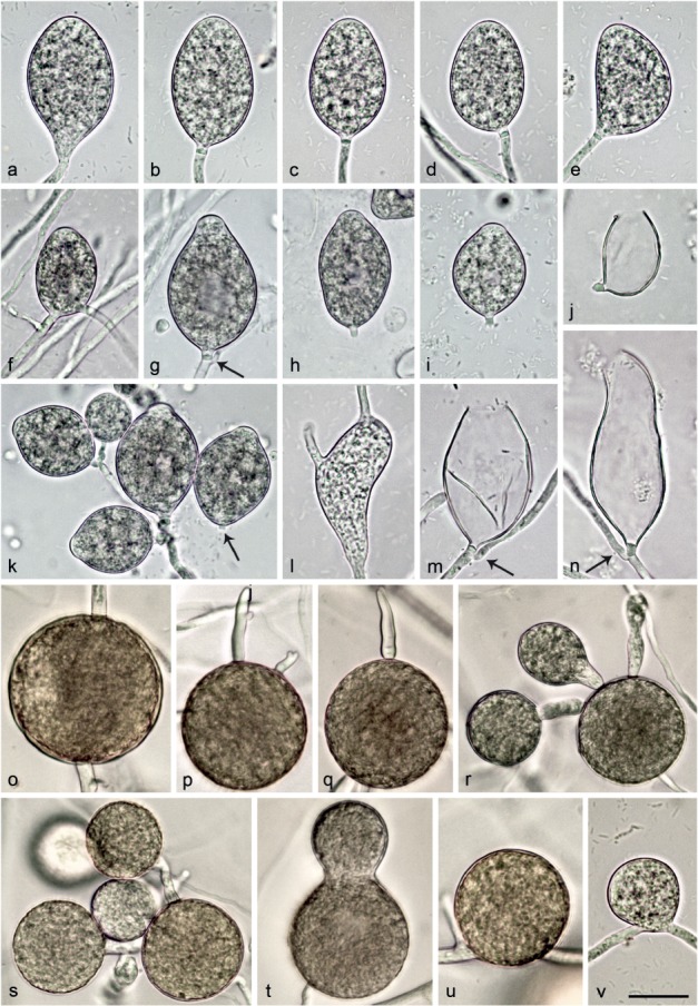
Fig. 6.

Morphological structures of Nothophytophthora chlamydospora. — a–n. Structures formed on V8 agar flooded with soil extract; a–i. mature non-papillate sporangia; a–e. borne terminally on unbranched sporangiophores; a. ellipsoid with tapering base; b–e. with conspicuous basal plugs; b. ellipsoid; c. ovoid; d. ovoid with slight lateral attachment of sporangiophore; e. mouse-shaped with laterally attached sporangiophore; f. ovoid, intercalary inserted; g. ovoid with vacuole, basal plug and beginning external proliferation (arrow); h–i. caducous with short pedicel-like basal plugs and small vacuoles; h. ellipsoid; i. limoniform; j. ovoid caducous sporangium with short pedicel-like basal plug, after release of zoospores; k. dense sympodium of ovoid to limoniform sporangia with shallow semipapillate apices; one sporangium caducous with short pedicel-like basal plug (arrow); l. sporangium which failed to form a basal septum and continued to grow at the apex, functionally becoming a hyphal swelling; m–n. empty sporangia after release of zoospores, with conspicuous basal plugs and external proliferation close to the base; m. ovoid; n. elongated-obpyriform with curved apex; o–v. structures formed in solid V8 agar; o–u. chlamydospores; o. globose, intercalary inserted; p–q. globose, terminally inserted with hyphal outgrowths; r–s. globose with radiating hyphae forming hyphal swellings or secondary chlamydospores; t. ampulliform, terminally inserted; u. globose, laterally sessile; v. intercalary globose hyphal swelling. — Scale bar = 25 μm, applies to a–v.
