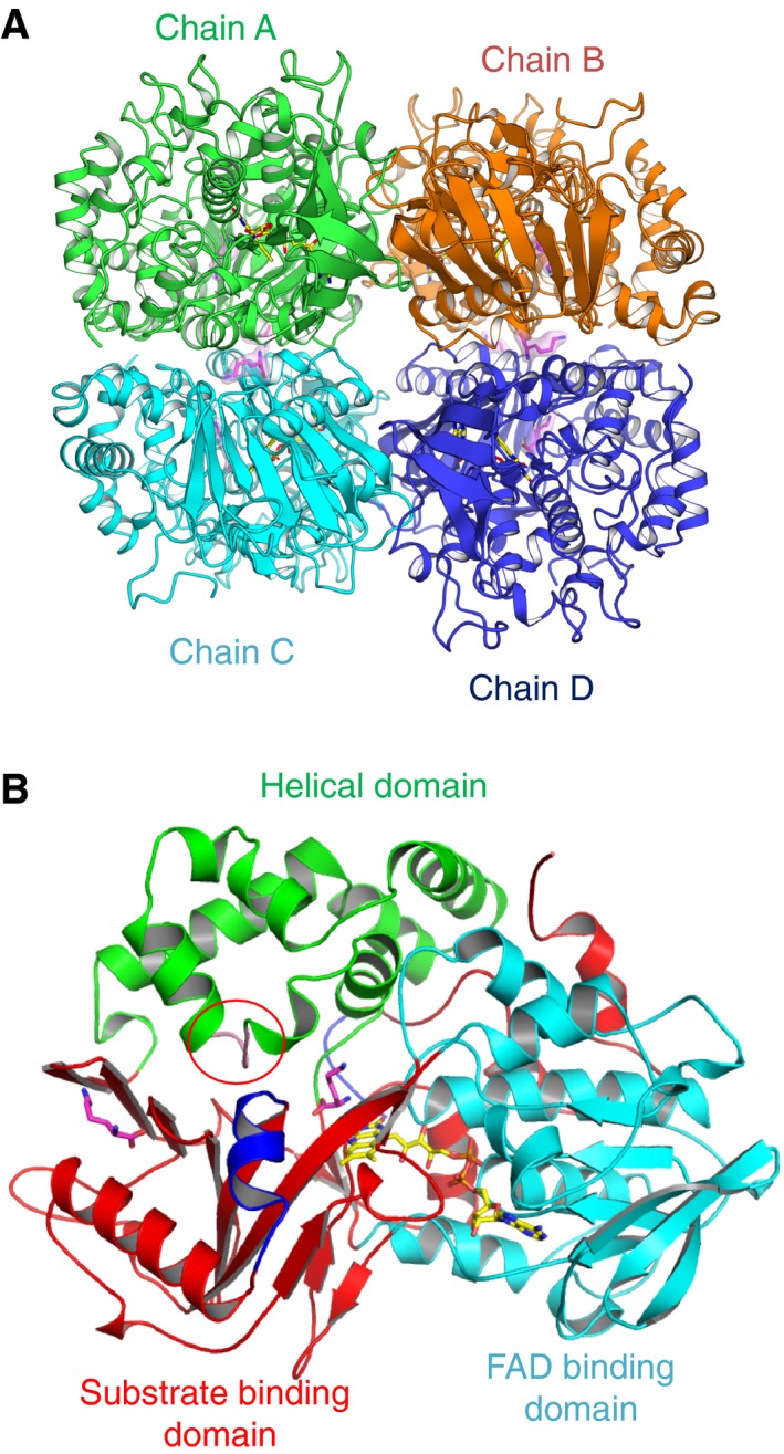
Figure 2.

Overall structure of l‐ AAO/MOG. (A) Tetramer structure contained in the asymmetric unit. FAD and l‐Lys molecules are shown as yellow and magenta sticks. The biological dimers consist of chains A–C and B–D. (B) Monomer structure of l‐ AAO/MOG. The plug loop (in the helical domain, red circle) and flexible helix (in the substrate binding domain) are shown in pink and blue, respectively.
