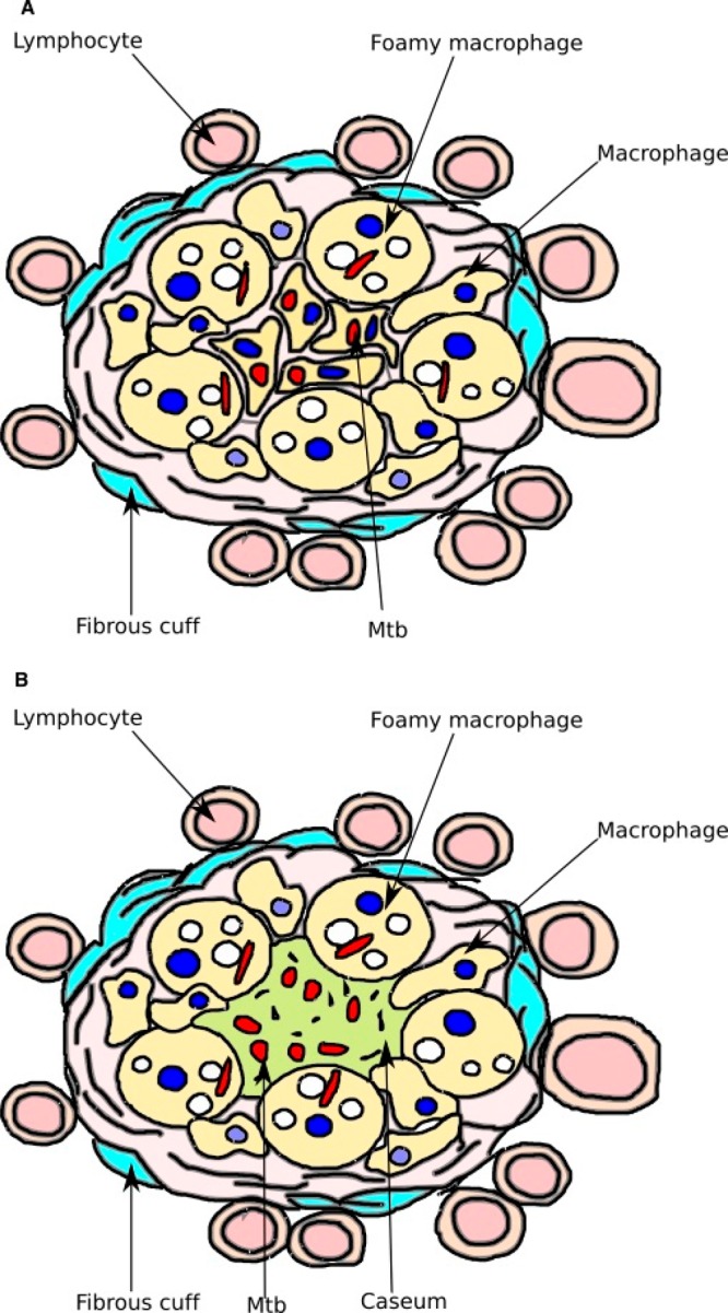
Figure 1.

Diagrams of characteristic granulomas. (A) Cellular granuloma. Macrophages infected with Mtb (red) are at the center. Lipid bodies (white) are within foamy macrophages. (B) Necrotic granuloma. Macrophages have died and released Mtb into the necrotic center, which is hypoxic and filled with lipid caseum.
