Figure 5.

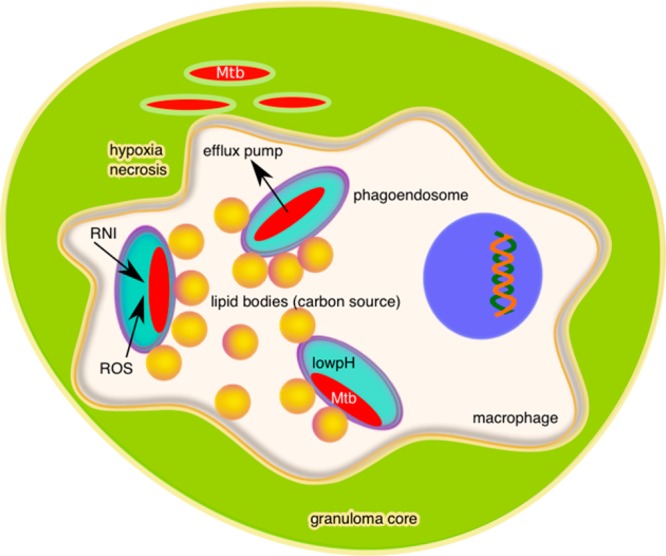
Diagram of an Mtb-infected macrophage within the necrotic core of a granuloma illustrating the intracellular and extracellular microenvironments which Mtb encounters.

Diagram of an Mtb-infected macrophage within the necrotic core of a granuloma illustrating the intracellular and extracellular microenvironments which Mtb encounters.