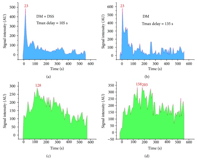
Figure 5.
A plot of signal intensity over time over the renal cortex and renal pelvis of the kidney. (a, c) The left shows the spectrum collected from a mouse in the DM + DSS group [peak at 23 s and 128 s in the renal cortex (a) and renal pelvis (c), respectively; Tmax delay = 105 s]. (b, d) The right shows the spectra collected from a mouse in the DM group [peak at 23 s and 158 s in the renal cortex (b) and renal pelvis (d), respectively; Tmax delay = 135 s].

