
Fig. 4.
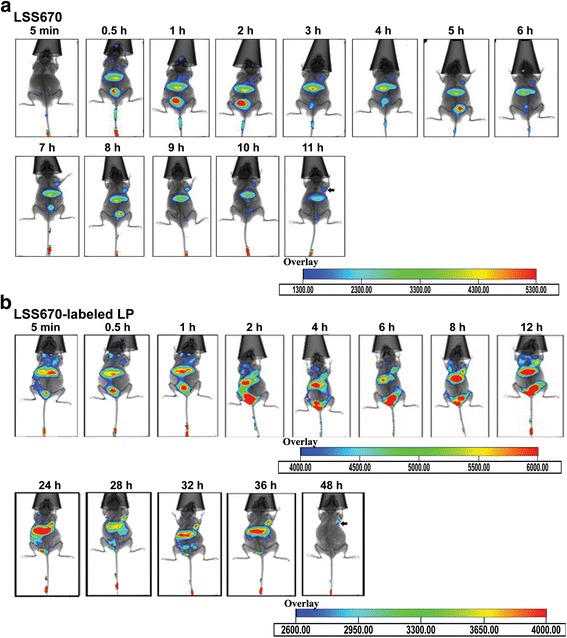
In vivo fluorescent images of nude mice bearing MDA-MB-231-Luc breast cancer xenografts implanted under the right front axilla. a After i.v. injection of free LSS670 dye. b i.v. injection of LSS670-labeled Lp. Fluorescent images were acquired from 5 min to 48 h after i.v. injection, n = 4. Optical imaging was performed with the Kodak in vivo FXPro imaging system, which combines multispectral fluorescence, luminescence, and digital X-ray capabilities in a single system. The excitation and emission filters were set at 650 and 700 nm, respectively