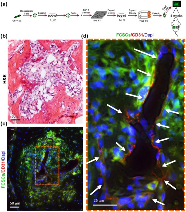
Figure 2.
A single fibrocartilage stem cell (FCSC) supports host blood vessel formation during transition from cartilage to bone in xenograft model at 4 wk. (a) Schematic showing single FCSC isolation. Heterogeneous GFP+ FCSCs were derived from the temporomandibular joint (TMJ) superficial zone (SZ) tissue from a GFP transgenic rat. GFP+ FCSCs were expanded in vitro and fluorescence-activated cell sorting (FACS) was used to plate a single cell/well into a 96-well plate. A total of 17 single-cell colonies were expanded over passages 2 to 3, seeded onto a collagen sponge, and surgically transplanted subcutaneously on the dorsum of nude mice for 4 wk. (b) Representative hematoxylin and eosin (H&E) stain of 2 of 17 xenografts at 4 wk showing transition to vascularized bone-like tissue at 4 wk. Scale bar = 50 µm. (c) Immunohistochemistry of CD31 (red) shows new blood vessel formation (arrows) surrounded by GFP+ FCSCs. (d) Orange dashed box is shown in higher magnification on the right. Scale bar = 50 µm and 25 µm, respectively.

