Figure 5.
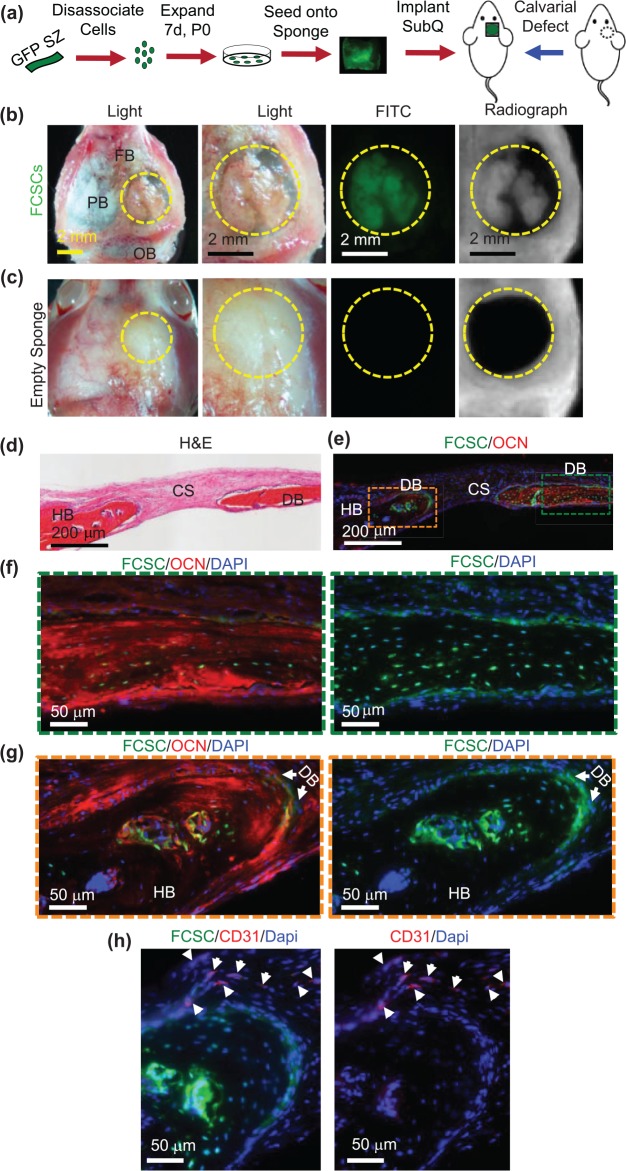
Transplanted GFP+ fibrocartilage stem cells engraft and repair calvarial bone defect. (a) Schematic of GFP+ fibrocartilage stem cell (FCSC) transplantation experiment in a mouse calvarial defect model. (b) Superior view of nude mouse parietal bone (PB) defect (defect = yellow dashed circle; FB, frontal bone; OB, occipital bone) with transplanted GFP+ FCSCs seeded onto a collagen sponge. FITC microscopy and radiographs demonstrated GFP+ FCSCs and radio-opaque tissue formed within transplants in the calvaria defect. (c) Superior view of the nude mouse calvarial bone defect model transplanted with empty sponge negative control. (d) Coronal hematoxylin and eosin (H&E) sections of FCSC xenografts (DB, de novo bone; HB, host bone; CS, collagen sponge). (e–g) Immunohistochemistry showed donor GFP+ FCSCs engrafted into a host calvarial bone defect and formed de novo bone (GFP+, osteocalcin+). Green = FCSCs; red = osteocalcin; blue = DAPI. (h) Immunohistochemistry showed host CD31+ endothelial cells localized at the periphery of newly formed bone. Green = FCSCs; red = CD31.

