Figure 1.
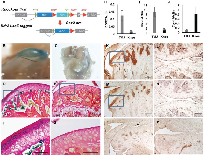
Discoidin domain receptor 2 (DDR2) is preferentially expressed and activated in temporomandibular joint (TMJ) articular fibrocartilage. (A) Strategy for developing Ddr2 LacZ-tagged allele mice. Mice with recombinant allele knocked into the Ddr2 locus (“knockout first” allele) were developed as described in the methods and crossed with global Cre (Sox2-Cre) mice to generate a germline Ddr2 LacZ-tagged allele (Ddr2+/LacZ). (B–G) LacZ localization in mandibular condyle (B, D, F) and knee joint (C, E, G) from 2-mo-old Ddr2+/LacZ mice. Whole mount LacZ staining (B, C). Low- (D, E) and high-magnification (F, G) histologic sections; high-magnification images are of boxed areas in D, E. (H–J) Reverse transcription quantitative real-time polymerase chain reaction detection of Ddr2 (H), Col1a1 (I), and Col2a1 (J) mRNA in TMJ and knee articular cells from 2-mo-old wild-type mice (n = 6). Values were normalized to β-actin mRNA. (K–P) Immunohistochemistry: TMJ (K, M, O, P) and knee joint (L, N). Antibodies: anti-total DDR2 (K, L, O) and anti-phospho-DDR2 (Y740; M, N, P). (O, P) To determine background staining, TMJs from Ddr2slie/slie mice were stained with total (O) and phosphor-DDR2 (P) antibodies. Scale bars: 0.2 mm in panel B; 0.5 mm for panel C; 40 µm for panels D, E, K–P; 20 µm for panels F, G. Arrow (O) indicates defects in condylar morphology of Ddr2slie/slie mice.

