Figure 2.

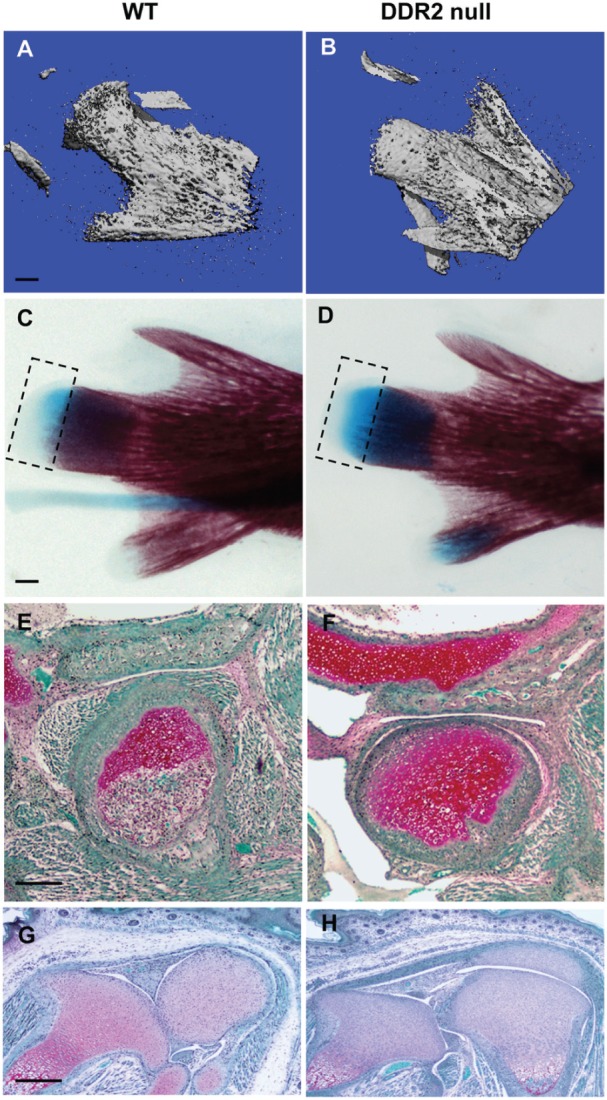
Ddr2-deficient mice exhibit tissue-selective defects in temporomandibular joint (TMJ) development. TMJ condyle structure was compared in newborn wild-type (WT) and Ddr2slie/slie mice with 3-dimensional reconstructed micro–computed tomography images (A, B), alizarin red and alcian blue whole mount staining (C, D), and safranin O staining of TMJ sections (E, F). Safranin O staining of knee joint histologic sections is also shown (G, H). Scale bars: 0.2 mm in panel A (A, B); 0.5 mm in panel C (C, D); 40 µm in panel E (E, F); 100 µm in panel G (G, H). Boxed area in panels C and D indicates alcian blue–stained region.
