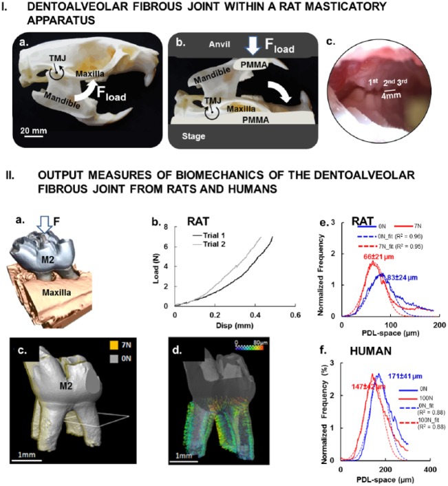
Figure 2.
Dentoalveolar fibrous joint within a rat masticatory apparatus. (I) Natural articulation was encouraged by maintaining the (a) rotational guidance about the temporomandibular joint of the masticatory complex and (b) loading (Fload) thereby interdigitation of maxillary and mandibular molars (zoomed view) (c) by loading a freshly dissected rat head with a mechanical testing device. (II) Output measures of biomechanics of the dentoalveolar fibrous joints from rats and humans. (a) Rat maxillary second molar was digitally isolated to determine the change in position of the tooth relative to the alveolar socket when loaded. (b) Load-displacement curves illustrate a nonlinear behavior of the dentoalveolar fibrous joint as the tooth is loaded and traverses into the alveolar socket of a rat. (c) Image registration of no-load (gray) and loaded (7 N, yellow) second molar of a rat allows evaluation of (d) surface distance representative of rat PDL deformation, which is illustrated by vectors (see scale bar). Histogram: distribution of PDL-space to normalized frequency at which a specific PDL-space occurs illustrates peak shifts as a result of tooth loading in rats (e) and humans (f), respectively. PDL, periodontal ligament.

