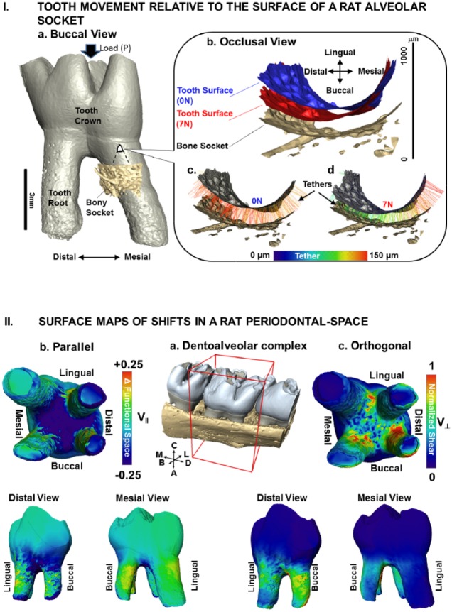
Figure 3.
Tooth movement relative to the surface of a rat alveolar socket. (I) (a) Volume-rendered 3-dimensional structure of a tooth relative to a portion of the alveolar bony socket. (b) A magnified region of the bony socket with positions of the tooth-root relative to the alveolar socket surface at no-load (0 N; c) and loaded (7 N; d) conditions illustrates colored tethers indicative of different lengths (see scale bar). (II) Surface maps of shifts in a rat periodontal space. (a) Surface-rendered 3-dimensional structure of molars in alveolar bone with the second molar for which V|| and V⊥ vectors were evaluated is highlighted in a box with global directions. (b) Components parallel to the tether were defined as narrowed or widened regions (compression and tension; V||) while (c) orthogonal components were defined as shear (V⊥) of the PDL and are illustrated as surface maps on the second molar. Widened PDL-space and less shear are seen in the mesial direction, while narrowing and more shear are seen in the distal direction. Global directions: A, apical; B, buccal; C, coronal; D, distal; L, lingual; M, mesial. PDL, periodontal ligament.

