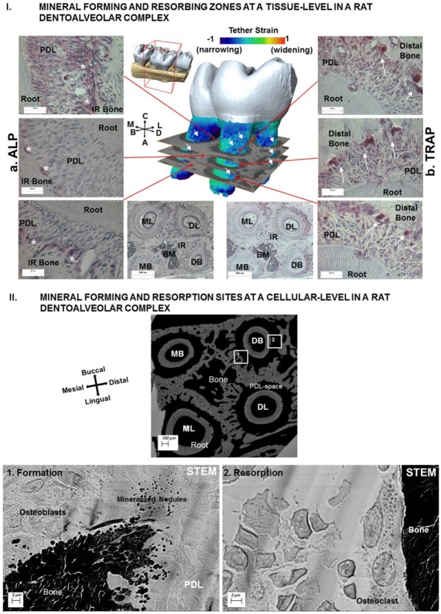
Figure 4.
Mineral-forming and mineral-resorbing zones at a tissue level in a rat dentoalveolar complex. (I) Spatial distribution of ALP and TRAP staining is shown in left (a) and right (b) panels: (a) ALP was mainly observed at the PDL-bone enthesis in the interradicular regions and within the interradicular endosteal spaces. (b) TRAP was observed at the PDL-bone enthesis on the distal regions of the complex. (II) Mineral-forming and mineral-resorption sites at a cellular level in a rat dentoalveolar complex. Morphologic features at an ultrastructural level between mineral-forming (1) regions of the second molar containing 4 roots are shown in the STEM micrograph. Higher-resolution micrographs of regions 1 and 2 illustrate mineralized nodules and osteoblasts at the PDL-bone enthesis. In region 2 opposite to the formation site (right image), the PDL-bone enthesis was smooth, and mineralized nodules were not observed. Instead, a multinucleated cell adhered to bone surface, representing an osteoclast responsible for active resorption, was observed. ALP, alkaline phosphatase; B, buccal; D, distal; L, lingual; M, mesial; PDL, periodontal ligament; STEM, scanning transmission electron microscope; TRAP, tartrate-resistant acid phosphatase.

