Figure 1.

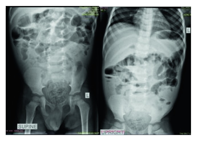
Plain abdominal X-rays (erect and supine) showing proximal small bowel loop dilatation, gaseous with some air fluid levels, while the distal part being dense with the rectum loaded with fecal matters.

Plain abdominal X-rays (erect and supine) showing proximal small bowel loop dilatation, gaseous with some air fluid levels, while the distal part being dense with the rectum loaded with fecal matters.